Each innominate bone is composed of three united bones. The anterior muscles posteriorly tilt the pelvis the posterior muscles anteriorly tilt the pelvis the muscles on the right side elevate the right side of the pelvis and therefore depress the left side of the pelvis and the muscles on the left side elevate the left side of the pelvis and therefore depress the right side of the pelvis.

Male Hip Bones And Ligaments Labeled Rear View On White Stock Photo Download Image Now Istock
From the quiz author.
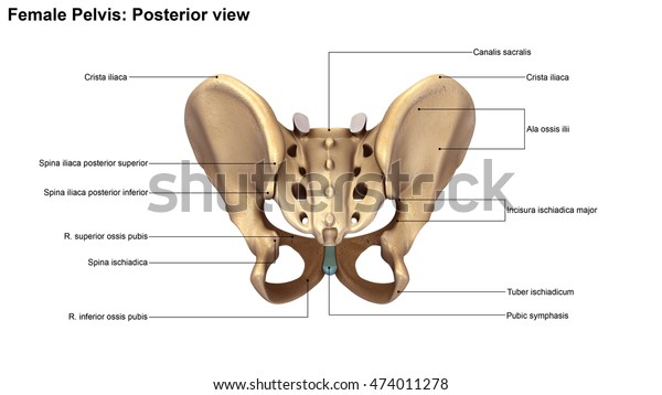
. The true pelvis or lesser pelvis lies below the pelvic brim Figure 1. Features that most clearly distinguish the female from the male pelvis include a wider subpubic angle wider sciatic notch and greater distance from pubic symphysis and anterior. Bony pelvis is formed posteriorly by the sacrum and the coccyx and laterally and.
We feature 65300000 royalty free photos 337000 stock footage clips digital videos vector clip art images clipart pictures background graphics medical illustrations and maps. Ilium ischium and pubis meeting in the acetabular fossa at the triradiate fusion. 182355869 stock photos online.
Trusted Medical Resource For Over 40 Years. Major components of the bony pelvis frontal superior view of the female pelvis. The pelvic spine is the posterior portion of the pelvis below the lumbar spine composed of the sacrum and coccyx.
COVID-FATE Related Anatomy Charts. Bony pelvis or pelvic skeleton is formed by hip bones sacrum and coccyx. The two pelvic bones are connected anteriorly by the pubic symphysis while posteriorly they articulate with the pelvic spine to form the sacroiliac joints.
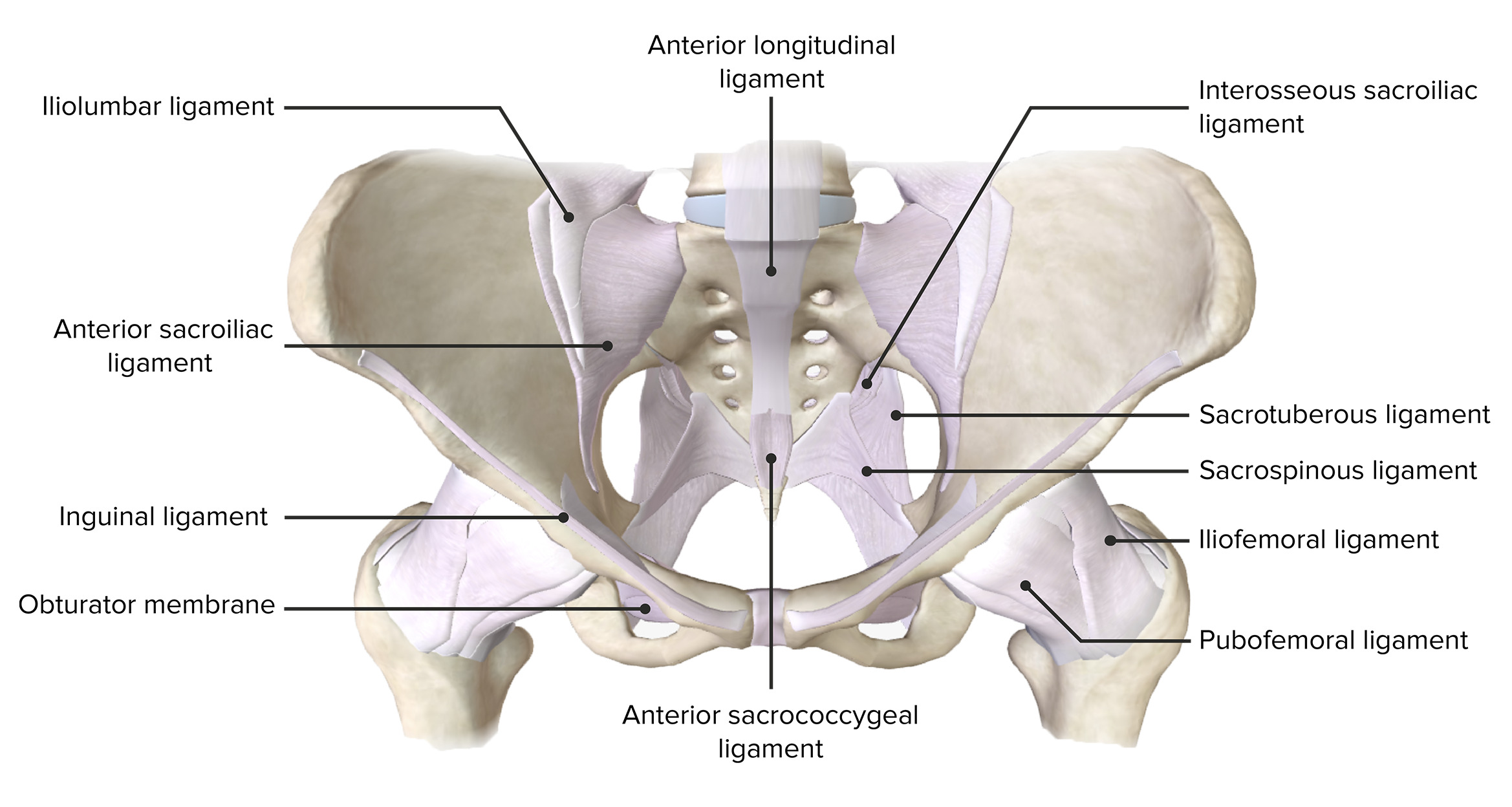
Download 1527 Posterior View Body Stock Illustrations Vectors Clipart for FREE or amazingly low rates. The three bones and three joints composing the pelvic ring have no inherent stability without vital ligamentous. Semimembranosus Flat muscle enabling the thigh to extend on the pelvis the knee to flex and the thigh and the leg to rotate inwardly toward the median axis.
Diagrams drawn by Miss Laura Hamilton. The pelvic region is the area between the trunk or main body and the lower extremities or legs. The female reproductive system is an intricate arrangement of structures that can separate into external and internal genitalia.
The sacrum and two innominate bones. This is an online quiz called THS Anatomy Pelvis Posterior View. There is a printable worksheet available for download here so you can take the quiz with pen and paper.
Click on the tags below to find other quizzes on the same subject. The anatomy of the pelvis is shown in Figure 1. The Adult Heart- Posterior Surface View.
The true pelvis situated inferior to the caudal portion of the parietal peritoneum is considered the pelvic cavity Figure 41-3 and Box 41-1 The posterior wall of the pelvic cavity is formed by the sacrum and coccyx and the margins of the posterolateral wall are formed by the piriformis and coccygeus muscles Figure 41-4. The male pelvis is different from a. A Anterior view of the hemipelvis.
The Adult Heart- Posterior Surface View. Bones of the Pelvis and Lower Back. There is a printable worksheet available for download here so you can take the quiz with pen and paper.
The pelvis consists of two hip bones attached at the front anterior by the pubic symphysis and at the back posterior by the sacrum. The vertebral column of the lower back includes the five lumbar vertebrae the sacrum and the coccyx. The external genitalia comprises the structures outside of the true pelvis including the labia majora and minora vestibule Bartholin glands Skene glands clitoris mons pubis perineum urethral meatus and periurethral area.
The pelvis is a ring structure made up of three bones. This landmark begins at the level of the sacral promontory posteriorly and the pubic symphysis anteriorly. The sacrum and two innominate bones.
Posterior view of the joints of the pelvis and hip. Stock Illustration - LifeART. The bones of the pelvis and lower back work together to support the bodys weight anchor the abdominal and hip muscles and protect the delicate vital organs of the vertebral and abdominopelvic cavities.
The pelvic cavity and perineum. The pelvic girdle or the bony pelvis is a bony ring formed by the left and right hip bones and the sacrum and it surrounds the pelvic cavity and connects the vertebral column to the lower limbs. The pelvis is a ring structure made up of three bones.
Anatomy of the pelvis. Share This Article. In females the pelvis also houses the uterus fallopian tubes and ovaries.
Abdominal. Each hip bone consists of an ilium ischium and pubis. The plane of the pelvic brim faces forward and forms an angle of about 60 degrees to the horizontal.
Pelvic anatomy is composed of two innominate coxal bones that articulate with the sacrum and proximal femora. Identify the following parts of the pelvic girdle This quiz has tags. And the thigh to extend on the pelvis.
This is an online quiz called Posterior view of Pelvic Anatomy SI ligaments. Ga305008 Fotosearch Stock Photography and Stock Footage helps you find the perfect photo or footage fast. The posterior wall is next to the perineal body rectum and peritoneal cavity at the pouch of Douglas while the two lateral walls lie against the pelvic diaphragm and major vaginal vessels.
B posterior view of hemipelvis. False and True Pelves. Pelvic briminlet - a line from the sacral promontory to the upper part of the pubic symphysis False pelvis - lies above this line Contains no pelvic organs except urinary bladder when full and uterus during pregnancy True pelvis - the bony pelvis inferior to the pelvic brim has an inlet an outlet and a cavity Pelvic axis - path of.
The main functions of the pelvic girdle are to transfer the weight of the upper body to the lower limbs when sitting or standing and provide attachment points for muscles that help with. Ad Great Prices on 10000 Products. Bony pelvis or pelvic skeleton is formed by hip bones sacrum and coccyx.
Female Pelvic Anatomy Charts. The three bones and three joints composing the pelvic ring have no inherent stability without vital ligamentous structures. The pelvis plays several important functions in the human body.
Anatomical landmarks within the vagina can be used to locate the position of such structures as the ureter and urethra and warn of their possible involvement in a vaginal laceration. Umbilical ligaments posterior view. 3d205010 Fotosearch Stock Photography and Stock Footage helps you find the perfect photo or footage fast.
We feature 66200000 royalty free photos 337000 stock footage clips digital videos vector clip art images clipart pictures background graphics medical illustrations and maps. New users enjoy 60 OFF. The space below contains the bladder rectum and part of the descending colon.
The pelvic region of the trunk is the lower part of the trunk between the abdomen and the thighs. Download high-res image 2MB Download.
Pelvis Anatomy Concise Medical Knowledge
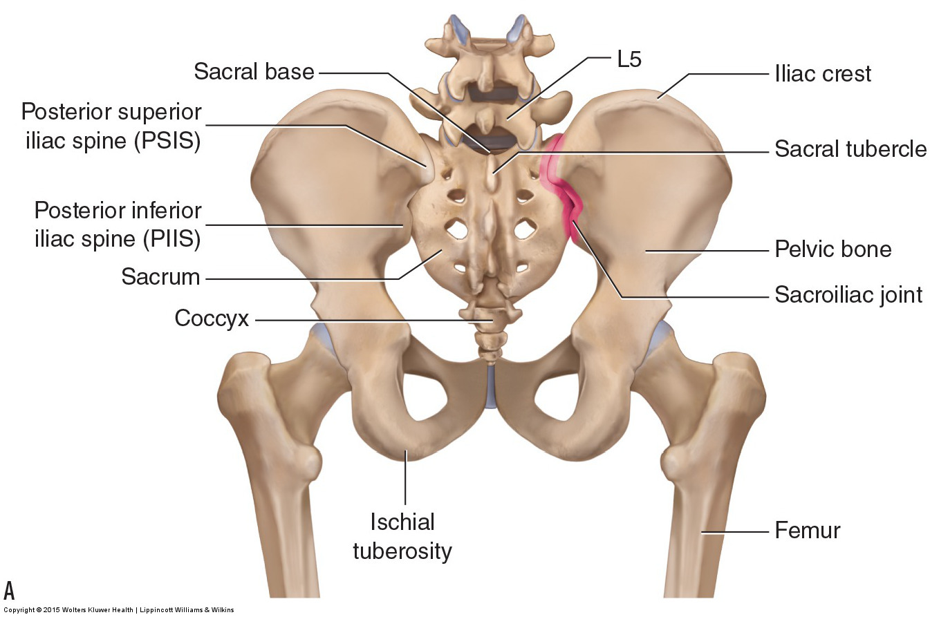
Bones Of The Lumbar Spine And Pelvis

The Pelvic Girdle And Pelvis Anatomy And Physiology I

Three Dimensional Posterior View Of The Pelvis Download Scientific Diagram
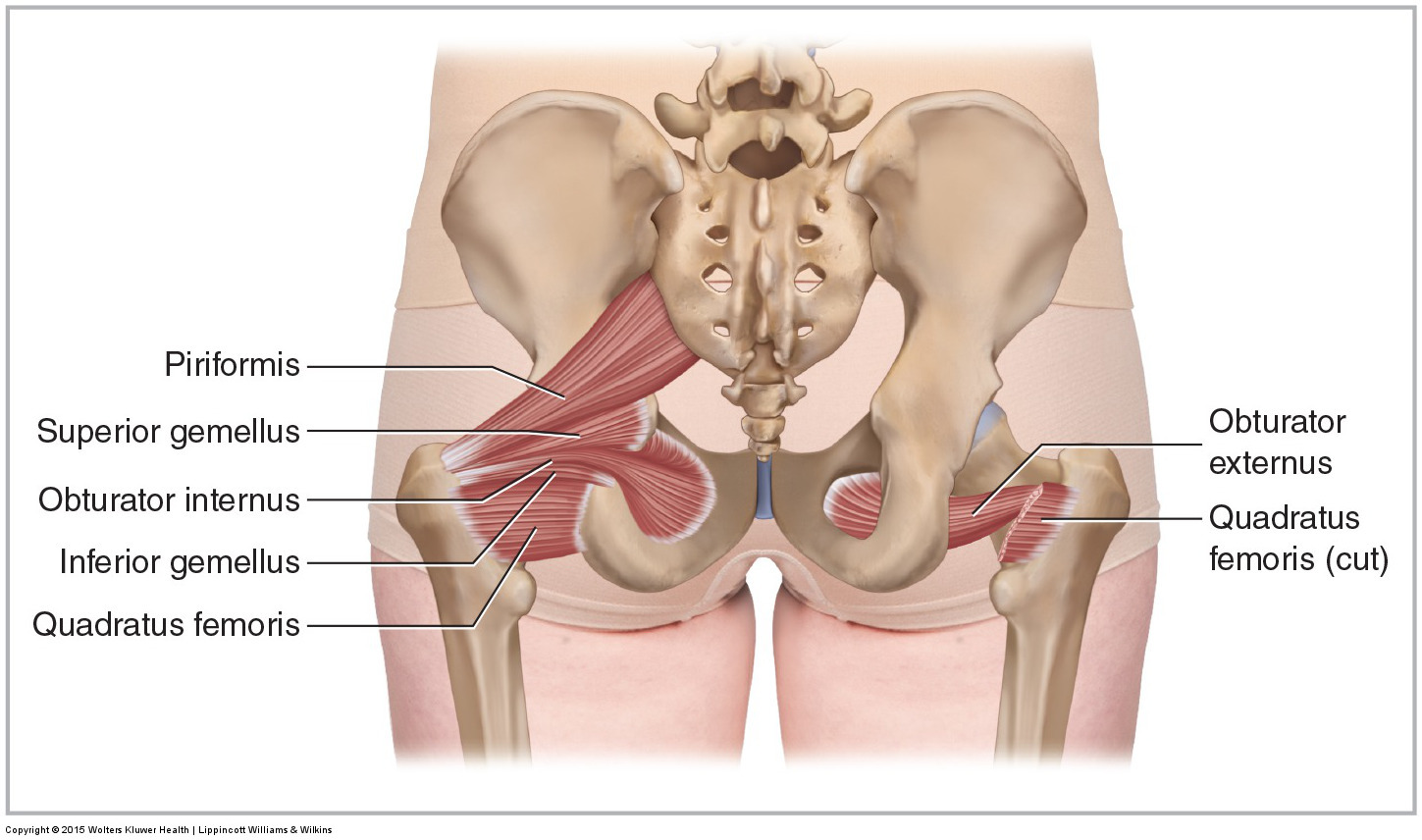
Muscles Of The Pelvis
Pelvis And Hip Anatomy Poster

Pelvis Anatomy Recon Orthobullets
Skeleton Pelvis Posterior View 3d Illustration Stock Illustration 474011278
0 comments
Post a Comment